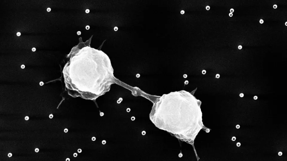
“Not far from the manners of Lennart Nilsson, Sara Davidsson Bencker has captured two pancreatic cells at the end of their cell division cycle, connected to the top of nanotubes. The image clearly visualizes both the cells and the nanowires and their interactions and communicates their size ratio and difference in magnitude. At the same time, the image also triggers associations of space and the universe and could illustrate the eternal struggle against loneliness.
This intriguing picture reminds us of the inherent contradiction between our perceived smallness on earth and the importance of yet contributing to the scientific community.”
This is the jury’s statement for awarding Sara the 1st prize in both categories. The prize consists of a wall-ready print of the photograph she submitted.
“That is great to hear, thank you very much! I’m very happy about the picture myself, and I think it fit nicely into my thesis on transfecting beta cells through nanotubes,” says Sara Davidsson Bencker when she receives the news.
The second and third prizes in each category are:
Category: “Best visual communication of scientific content”
2nd Prize: Quantum Ring
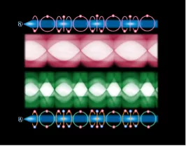
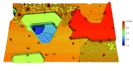
We see here how Claes Thelander has created a pattern of a quantum ring fabricated inside a quantum dot, rotated relative to a magnetic field. It is probed by electron tunneling spectroscopy that senses excited states, which appear as a change in color contrast. Almost as if Marimekko was involved in the design, the image illustrates clearly and in an aesthetically appealing way both the scientific data and the underlying physics.
3rd Prize: Looking through microspheres
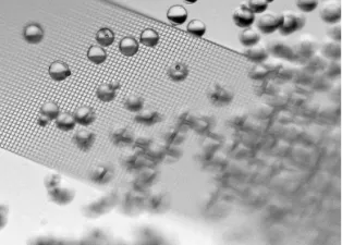
Jason Beech takes us back to the Golden Age of Dutch science and technology, when microbiologist Antonie van Leeuwenhoek in the early 1700’s looked through a sphere of glass and discovered that we are surrounded by uncountable living things:
“With a glass bead a few mm in diameter, some ingenuity, and patience, a human being discovered that we are part of a web of life more diverse and numerous than we had ever imagined.”
The magnification of a glass bead is related to its diameter, through the so-called focal length. While van Leeuwenhoek used millimeter-sized beads and could observe microbes around a micrometer size, these micrometer beads allow us to see the structures of a microfluidics device directly underneath them. We would love to hear more stories like this!
Category: “Most aesthetically appealing image”
2nd Prize: Fluorescent Nanoflower

Sometimes, defects catch our eyes not by their imperfection but by their beauty. In this picture, we see Julia Valderas Gutiérrez’ supported lipid bilayers – complex fluid systems similar to cell membranes and composed of mobile lipids that play a crucial role on the cell’s survival. Model membranes can be formed and studied on vertical arrays of light-guiding silicon nanowires adding lipid vesicles that fuse together when a critical concentration is reached. With epifluorescence microscopy, she shows us a wave effect suddenly “engulfing” the nanowires, and the lipids, whose fluorescence is enhanced by the nanowires, diffuse laterally within the membrane. Most of all, we liked the fluid membrane flowing around this flower-like beautiful defect of the nanowire platform. The image will continue to fascinate an audience beyond the scientific community – and we chose to make it the cover of the NanoLund Annual Report.
3rd Prize: Storm-coloured Noise
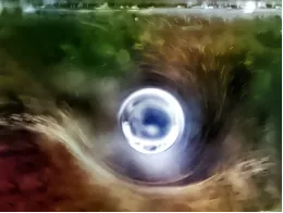
A distinct example of when laboratory work has slightly gone south but instead generated imaginative pictures of the nano-universe. This air bubble got stuck inside the microfluidic channel in a microscope picture of an acoustofluidic chip – consisting of a piezoelectric element attached to a silicon die with an etched microchannel sealed by a glass wafer, during operation. Like in an underwater action film, we see 5 µm big fluorescent polystyrene particles immersed in water, focused by acoustic forces toward the centre of the channel but on their way through the device, deflected by the vibrating bubble. A usually highly undesired scenario that in this case leads to a stunning scene worth capturing – by Michael Gerlt.
Special mentions
During our work choosing the winners of this competition, we couldn’t help but develop our personal favourites. Brace yourselves for our Special mentions!
Special mention: Steam Punk / Nostalgia
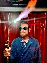
Filip Lenrick, photographed by Klas Holger Jönsson, wearing 1930s welding goggles and a welding robe inscribed with “Mekanisk Teknologi och Verktygsmaskiner” sets an example for the type of staff photo we would love to see more of!
As he puts it: “Bridging the gap between traditional mechanical technology and the realm of nanotechnology”.
Special mention: Brutalism
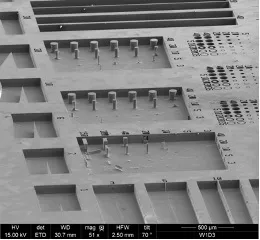
Michael Gerlt is, contrary to what you might think, not a brutalist architect from the 1950s. This picture shows various structures etched to a depth of 100 µm in a process optimized to lead to an etch angle very close to 90° allowing to etch a wall between two microfluidic channels as thin as 10 µm. If the structure is too thin, it simply falls down as can be nicely seen at the section containing rectangular structures. We like how Michael clearly shows what is being done, the scale, the difficulty, and the limits of what is possible today.
Special mention: Fantasy / Imagination

This one really sparks the imagination. Anastasia Ttsioki’s lovely artwork of snail antennas and turtles illustrates heteroepitaxial growth of materials. It has been featured on the cover of a scientific journal, and it could perfectly well hang on the wall in the foyer of Fysicum as well as in a primary school or physics entrance hall.
Special mention: Minecraft
Rohit Yadav’s picture “Atomic growth on (111) islands” brings out our inner child and we are positive that this atomic investigation of metal-semiconductor interface is interesting not only for fundamental science, but also for inspiring and attracting the gaming generation into the labs.
Special mention: UFO
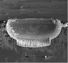
What is it? Pests? A space ship? A jellyfish? A universalistic dystopia? No, it’s Michael Gerlt, sharing his inductively coupled plasma deep reactive iron etching with us. When the process parameters may not be optimized, the result can be: black silicon. We love this and are so thankful for the generosity in showing scientific results that might not be exactly what you wished for.
The jury, consisting of Anna Stenstam (chair), Heiner Linke, Maximilian Nitsch, and Evelina Lindén, wants to thank all of you for your contributions and for your patience. We had a difficult time picking the winners and you overwhelmed us with your fantastic work and innovative way of looking at the world.